Digital Photography
-
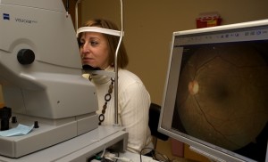
A patient has a digital photograph taken of her eye.
Fundus Photographs – pictures of specific areas in the back of the eye or of the optic nerve.
Slit Lamp Photographs – pictures of specific areas on the eyelids or the front part of the eye.
Fluorescein Angiography – a procedure which involves the dilation of your eye in order for a technician to take photographs of the back of your eye. After the dilation a dye is administered intravenously through your arm or hand. Photographs are taken of the retina as the dye circulates through the blood vessels of the eye. This helps detect any leaks or blockages of the blood vessels and determine the exact location of the problem.
Visual Fields
-
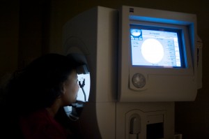
A visual field tests a patient’s peripheral vision.
Humphrey Visual Field – Automated visual field test measure side (peripheral) vision. This test will assist your doctor to detect any loss of side vision which can be a sign of glaucoma or can detect neurologic abnormalities.
Goldmann Visual Field – Manual visual fields performed by technicians to measure peripheral vision. This test will assist your doctor to detect any loss of side vision which can be a sign of glaucoma.
Ocular Ultrasounds – This test can capture two or three dimensional images of the eye.
B-Scan – ultrasound of the back of eye which can detect any abnormalities in the back of the eye including tumors, bleeding, swelling, foreign bodies or retinal detachment.
A-Scan – ultrasound which measures the eye to determine the proper power of the synthetic lens implant that will be used during cataract surgery.
UBM – ultrasound image of the front of the eye (anterior chamber) to detect evaluate anterior chamber angle as well as look for tumors, foreign bodies.
OCT (Optical Coherence Tomography)
-
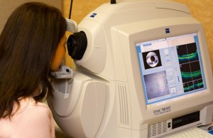
A patient is tested with Optical Coherence Tomography
OCT is a painless noninvasive technique which shows the thickness of the retinal layers of the eye. Your eyes are dilated in order to accurately view the back of the eye. This is used for diagnosing early Glaucoma, Macular Degeneration, and other retinal diseases.
Pachymetry – Measures the thickness of the cornea.
Corneal Topography – Measures the shape of the eye and any irregular surfaces of the cornea.
Electroretinogram (ERG) – Measures electrical pulses from tissues in the back of the eye to test visual function.


